Comprehensive Strategies for Detecting and Diagnosing Lung Cancer
This comprehensive article explores the various diagnostic approaches for lung cancer, emphasizing the importance of imaging techniques like X-ray, CT, MRI, and PET scans, alongside invasive biopsy procedures such as bronchoscopy and needle biopsy. Early detection through these methods significantly impacts treatment success and patient survival. The article also details staging procedures crucial for tailoring treatment strategies, ultimately facilitating better management and prognosis of lung carcinoma. A thorough understanding of these diagnostic tools ensures timely and accurate lung cancer diagnosis.
Comprehensive Strategies for Detecting and Diagnosing Lung Cancer
Lung cancer remains one of the most prevalent and deadly forms of cancer worldwide. Early detection and precise diagnosis are crucial steps in improving patient outcomes and increasing survival rates. Modern medical advancements have introduced a wide array of diagnostic techniques, including sophisticated imaging modalities and invasive procedures, enabling healthcare professionals to accurately identify and stage lung cancer. This comprehensive overview explores the critical approaches used to detect and diagnose lung carcinoma, highlighting the importance of each method in the diagnostic process.
Typically, the diagnostic journey begins with non-invasive imaging tests designed to detect abnormalities within the lungs. These initial assessments often reveal suspicious masses or nodules that warrant further investigation. Sometimes, simple sputum analysis under a microscope can uncover malignant cells, especially in early or accessible cases. However, further tests are generally needed to confirm a diagnosis, determine the exact nature of the abnormality, and establish the stage of the disease.
Understanding the combination of imaging techniques and invasive diagnostic procedures is essential in the effective management of lung cancer. This article discusses the most common diagnostic tools, including their indications, advantages, and limitations, providing a comprehensive overview of how lung cancer is identified, confirmed, and staged.
Initial Detection: The Role of Imaging Modalities
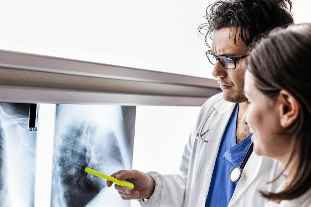
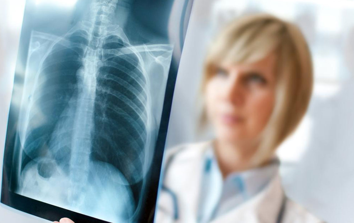
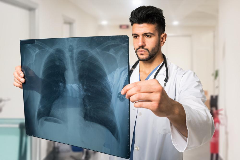
Imaging techniques are fundamental to the initial detection of lung anomalies. The most common starting point is a chest X-ray, a quick and accessible test that can reveal large masses, pleural effusions, or other abnormalities. However, chest X-rays may miss small or early-stage lesions due to their limited resolution.
To improve detection accuracy, computed tomography (CT) scans are often employed. CT scans provide detailed cross-sectional images of the lungs, allowing clinicians to identify small nodules or lesions that might be undetectable on an X-ray. Multi-detector CT technology has significantly enhanced the ability to detect early-stage tumors, assess their size and location, and plan further interventions.
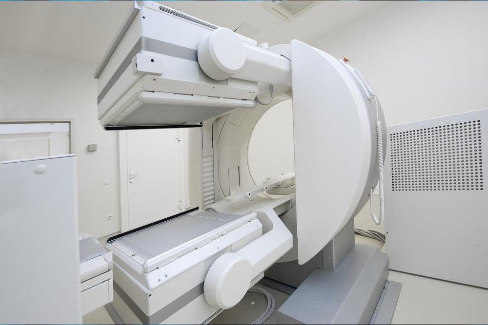
Beyond CT scans, advanced imaging modalities such as Magnetic Resonance Imaging (MRI) and Positron Emission Tomography (PET) scans are utilized for comprehensive evaluation. MRI offers superior soft tissue contrast, which can be helpful in assessing tumor invasion into surrounding structures. PET scans, often combined with CT (PET-CT), allow for metabolic imaging to distinguish benign from malignant lesions and detect potential metastases at an early stage.
Bone scans may be performed when metastasis to skeletal tissues is suspected, providing crucial information for staging and treatment planning.
Pathological Confirmation: Biopsy Techniques
While imaging can strongly suggest the presence of lung cancer, definitive diagnosis requires pathological confirmation. This is achieved through tissue sampling, or biopsy, which involves extracting a small piece of lung tissue for microscopic examination. Several biopsy techniques are available, tailored to the location and accessibility of the suspicious lesion.
Sputum Cytology: This non-invasive method involves examining sputum samples under a microscope for malignant cells. It is more effective in central tumors or those shedding malignant cells into the airways but has limitations in sensitivity.
Needle Biopsy: Percutaneous needle biopsy, often guided by CT imaging, allows sampling of suspicious lesions located on the outer parts of the lung. It provides high diagnostic accuracy with minimal invasiveness.
Bronchoscopy: This procedure involves inserting a flexible tube with a camera into the airways to visualize the bronchial tubes directly. During bronchoscopy, tissue samples can be taken from larger central tumors or lymph nodes.
Thoracoscopy and Video-Assisted Thoracic Surgery (VATS): For peripheral lesions or when other methods are inconclusive, minimally invasive surgical procedures such as thoracoscopy can obtain larger tissue samples, facilitating a more accurate diagnosis.
Staging and Assessing Disease Extent
Once lung cancer is diagnosed, accurate staging is crucial for selecting appropriate treatment strategies and predicting prognosis. Additional diagnostic tests are employed to evaluate if and where the cancer has spread beyond the lung. These include:
Endobronchial Ultrasound (EBUS): An minimally invasive procedure using ultrasound-guided bronchoscopy to assess lymph nodes within the mediastinum and determine metastasis.
Endoscopic Esophageal Ultrasound: Similar to EBUS, this technique provides detailed visualization of mediastinal structures and lymph nodes.
Mediastinoscopy: A surgical procedure allowing direct visualization and biopsy of mediastinal lymph nodes through a small incision at the base of the neck.
Mediastinotomy and Thoracoscopy: More invasive procedures used when extensive sampling of mediastinal tissues is required.
Additional Imaging for Metastasis: Bone scans, abdominal ultrasounds, or MRI scans may be performed to detect distant metastases, such as in the liver or bones.
Comprehensive staging informs the treatment approach, whether surgical resection, chemotherapy, radiation therapy, or targeted therapies.
In summary, detecting and diagnosing lung cancer involves a combination of imaging procedures for initial assessment and invasive biopsy techniques for definitive confirmation. The choice of diagnostic methods depends on tumor location, patient health, and overall clinical suspicion. An accurate diagnosis followed by precise staging is vital for developing an effective treatment plan and improving patient outcomes.