Comprehensive Guide to How MRI Scanners Operate and Their Role in Medical Diagnostics
This comprehensive guide explores how MRI scanners operate, including preparation steps, working principles, and their critical role in medical diagnostics. Detailed insights into the imaging process, safety considerations, and advancements highlight MRI’s importance in detecting internal health conditions with high precision. Suitable for patients and healthcare professionals alike, this article underscores MRI technology’s safety, accuracy, and evolving capabilities in modern medicine.
Understanding the Functionality of MRI Technology in Medical Imaging
Magnetic Resonance Imaging (MRI) has revolutionized diagnostic medicine by providing a non-invasive, highly detailed view of the internal structures of the human body. Unlike X-rays or CT scans, MRI employs strong magnetic fields combined with radiofrequency waves to produce clear, high-resolution images of tissues and organs. This capability is especially valuable for diagnosing and monitoring conditions involving soft tissues such as muscles, ligaments, cartilage, and internal organs. Its safety profile, primarily because it does not use ionizing radiation, makes MRI a preferred choice in many clinical scenarios.
The importance of MRI technology lies in its ability to deliver precise images that help healthcare professionals assess injuries, diseases, tumors, and other medical conditions accurately. It provides crucial information that informs treatment plans, including surgical interventions when necessary. Moreover, MRI scans can be tailored with specific contrast agents to highlight particular tissues or abnormalities, enhancing diagnostic accuracy.
Preparation Steps for an MRI Scan
Wear loose, metal-free clothing or a hospital gown as directed by your healthcare provider. This reduces interference with the magnetic field and ensures safety during the scan.
Inform your medical team if you have any allergies, especially to contrast dyes such as iodine or gadolinium, which might be administered to improve image clarity.
Discuss any medical implants, pacemakers, or metal fragments in your body, as these can affect the MRI procedure or pose safety risks.
In some cases, your doctor may schedule a contrast-enhanced MRI, where a contrast agent is injected into your vein to improve the visibility of certain tissues or blood vessels. This process enhances the diagnostic value of the MRI images, especially in detecting tumors, inflammation, or vascular abnormalities.
How MRI Scanners Function
The core principle of MRI involves aligning hydrogen protons—abundant in water and fat—within the body's tissues using a powerful magnetic field. When a radiofrequency pulse is applied, these protons are temporarily knocked out of alignment. As they realign with the magnetic field, they emit radio waves that are captured by sensitive detectors. Variations in the emitted signals correspond to different tissue densities and compositions, allowing the creation of detailed cross-sectional images.
This process enables MRI to differentiate among various types of tissues, providing excellent contrast between healthy and abnormal areas. Unlike X-ray or CT imaging, MRI doesn’t rely on radiation exposure but instead utilizes magnetic and radiofrequency energy, making it safer for repeated use and suitable for vulnerable populations such as pregnant women and children.
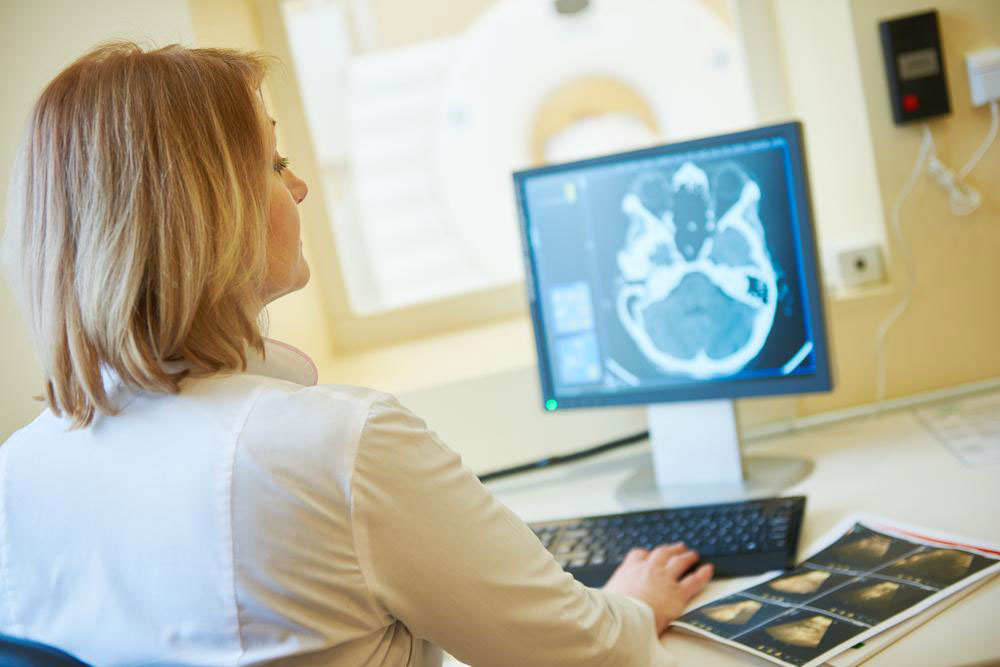
These emitted signals are processed by advanced computer algorithms to generate images. The images can be viewed from multiple angles and slices, aiding physicians in pinpointing lesions, tears, tumors, or other abnormalities with high precision. The superior soft tissue contrast of MRI makes it invaluable in detecting conditions that other imaging modalities might miss or overlook.
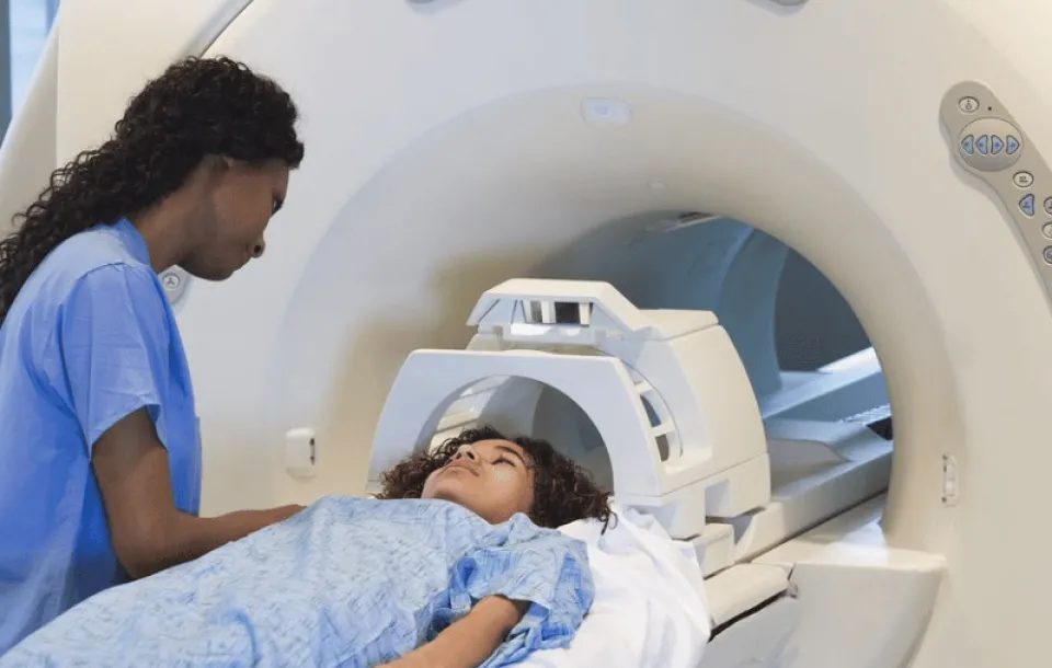
The MRI Examination Process for Knee Injuries and Beyond
The MRI procedure for the knee or other regions usually involves several steps to ensure both accuracy and patient comfort. Patients are typically positioned on a motorized table that slides into the MRI scanner. To prevent movement during imaging, straps or cushions are used to stabilize the limb. Small radio frequency coils—devices that detect the signals emitted by hydrogen nuclei—are placed around or near the area being scanned, substantially improving the clarity and resolution of the images.
The entire process generally lasts between 15 to 45 minutes, depending on the complexity of the examination and the area being studied. Most patients find MRI scans painless, although some may experience discomfort or anxiety, particularly if they have claustrophobia or fear enclosed spaces. In such cases, mild sedation might be offered to facilitate the procedure.
Post-scan, images are analyzed by radiologists, who interpret the findings and communicate the results to your healthcare provider. This comprehensive imaging capability has made MRI a crucial tool in diagnosing complex orthopedic injuries, neurological conditions, cancer, and many other health concerns.
With ongoing advancements in MRI technology—such as faster imaging sequences, higher magnetic field strengths, and functional MRI—this modality continues to evolve, offering even more precise and quicker diagnostic options for patients worldwide.