Comprehensive Guide to Knee MRI Imaging: What You Need to Know
This comprehensive guide explores knee MRI imaging, highlighting its importance in diagnosing knee injuries and conditions. It discusses how MRI works, what conditions it can detect, benefits, risks, and preparation tips. Suitable for patients seeking detailed understanding, it emphasizes the role of MRI in modern orthopedics, ensuring informed healthcare decisions for optimal knee health.

Comprehensive Guide to Knee MRI Imaging: What You Need to Know
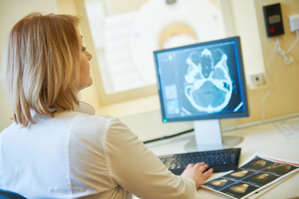
Magnetic Resonance Imaging (MRI) has revolutionized medical diagnostics by providing highly detailed images of the body's internal structures without the need for invasive procedures. Specifically, for knee health assessment, MRI offers invaluable insights that aid physicians in diagnosing a wide range of conditions. This detailed imaging technique is often preferred due to its non-invasive nature and superior clarity compared to other diagnostic tools. Understanding how knee MRI scans work, their benefits, potential risks, and preparation tips can empower patients to make informed decisions regarding their healthcare.
MRI technology relies on creating detailed cross-sectional images through the use of strong magnetic fields and radiofrequency waves. These images allow medical professionals to visualize bones, cartilage, tendons, muscles, blood vessels, and ligaments with remarkable precision. In the context of knee health, MRI plays a crucial role in identifying injuries, degenerative diseases, and other abnormalities that might not be apparent through physical examination or less detailed imaging modalities like X-rays.
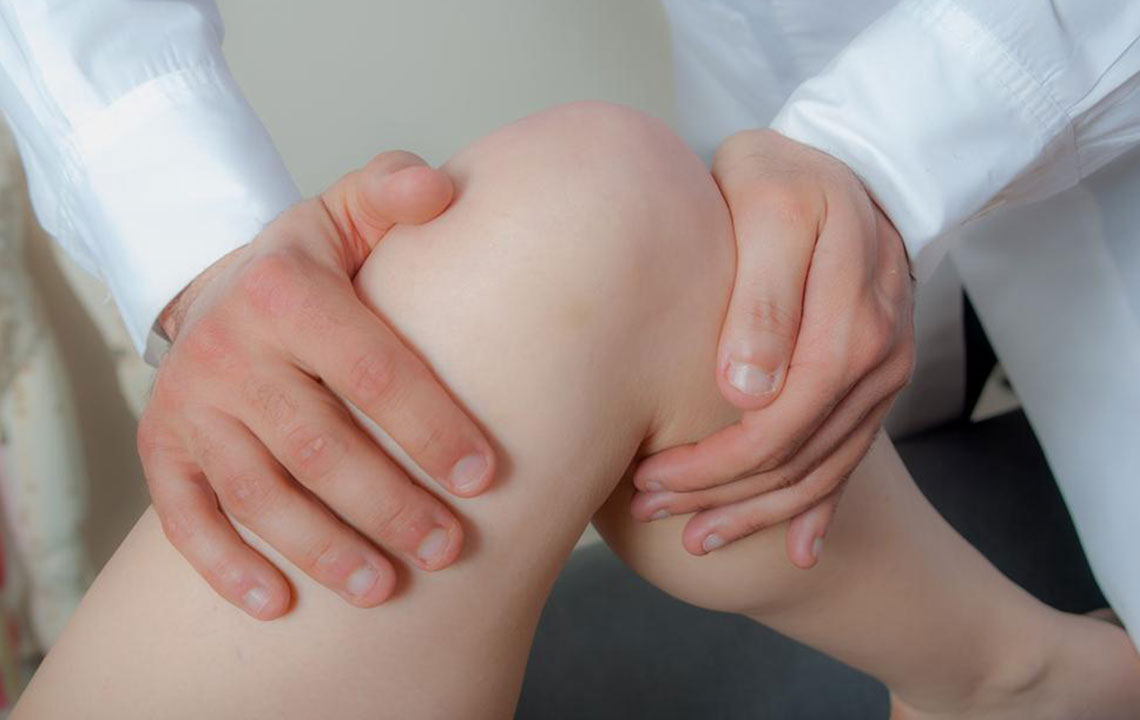
Doctors recommend knee MRI scans for various reasons, including persistent joint pain, swelling, instability, or if previous treatments haven't resolved symptoms. This imaging is especially useful for detecting ligament or cartilage tears, meniscal injuries, osteoarthritis progression, joint effusions, and even complex fractures that involve soft tissues. Moreover, MRI can assist in planning surgeries, monitoring recovery, and assessing post-operative complications, making it an indispensable tool in modern orthopedic diagnostics.
Aside from injury assessment, MRI is beneficial in diagnosing other conditions, such as tumors, infections, or blood vessel abnormalities around the knee. For athletes, MRI can help identify subtle sports injuries that might otherwise go undetected, enabling targeted treatments and quicker return to activity. Additionally, MRI scans can sometimes detect issues related to implanted medical devices, provided the necessary precautions are taken.
When doctors suspect degenerative joint diseases like osteoarthritis or rheumatoid arthritis, MRI offers detailed tissue visualization that can help determine disease severity and guide treatment plans. In cases where patients experience unexplained swelling or fluid accumulation, an MRI can identify the source of inflammation or bleeding. Furthermore, MRI is sometimes used in conjunction with other diagnostic procedures such as X-rays, ultrasound, or arthroscopy to provide a comprehensive assessment.
Although MRI is generally safe, understanding the potential risks and precautions is important. These include:
Metallic implants, such as pacemakers or certain orthopedic hardware, may interfere with MRI magnets, resulting in image distortion or device malfunction. Always notify your doctor about any implants or metal fragments.
Gadolinium-based contrast agents are sometimes used to enhance image clarity. In rare cases, these can cause allergic reactions or be linked to conditions like Nephrogenic Systemic Fibrosis (NSF) in patients with impaired kidney function.
Pregnant women are advised to postpone MRI scans unless absolutely necessary. If a scan is required, discuss potential risks and benefits with your healthcare provider.
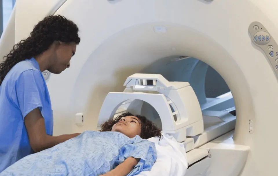
Prior to your MRI appointment, ensure you communicate fully with your medical team about any allergies, recent surgeries, or existing health conditions. Follow all pre-scan instructions carefully, including fasting if advised, and refrain from wearing jewelry or metal accessories. It's also worthwhile to inform the technician if you experience claustrophobia or anxiety, as arrangements such as sedation or open MRI alternatives might be available to improve comfort during the procedure.
In conclusion, knee MRI imaging is a powerful diagnostic tool that greatly enhances our ability to understand and treat a myriad of knee conditions. Its non-invasive nature, combined with high-resolution imaging capabilities, makes it the preferred choice for comprehensive knee assessment. Staying informed about the procedure, potential risks, and preparation steps can help patients undergo the process smoothly and effectively, ensuring the best possible outcomes for knee health management.