Comprehensive Guide to Minimally Invasive Ureteroscopy for Kidney Stone Treatment
Ureteroscopy has revolutionized kidney stone treatment by offering a minimally invasive, highly effective procedure to remove or break down stones. This article provides an in-depth overview of the procedure, from preparation to recovery, emphasizing its safety, success rate, and benefits. Discover how urologists use advanced technology like laser fragmentation and real-time imaging to ensure optimal patient outcomes, minimize complications, and promote rapid recovery. Whether dealing with small or large stones, ureteroscopy remains a cornerstone technique in modern urological care, providing relief and restoring health with precision and ease.

Comprehensive Guide to Minimally Invasive Ureteroscopy for Kidney Stone Treatment
The human kidneys play a crucial role in maintaining overall health by filtering waste products and excess fluids from the bloodstream, which are then excreted through urine. While the kidneys efficiently perform this vital function, certain chemicals and minerals in the body can crystallize within the renal system, leading to the formation of kidney stones. These stones vary greatly in size, shape, and composition, and their presence can cause significant discomfort, urinary obstruction, or infection if not appropriately managed.
Kidney stones are a common health concern affecting millions worldwide. The approach to treatment depends largely on the size, location, and type of the stone. Small stones, typically less than 5 millimeters in diameter, often pass naturally through the urinary tract with supportive care and medication. However, larger stones or those causing severe symptoms often require surgical intervention, with minimally invasive procedures like ureteroscopy being among the most effective solutions today.
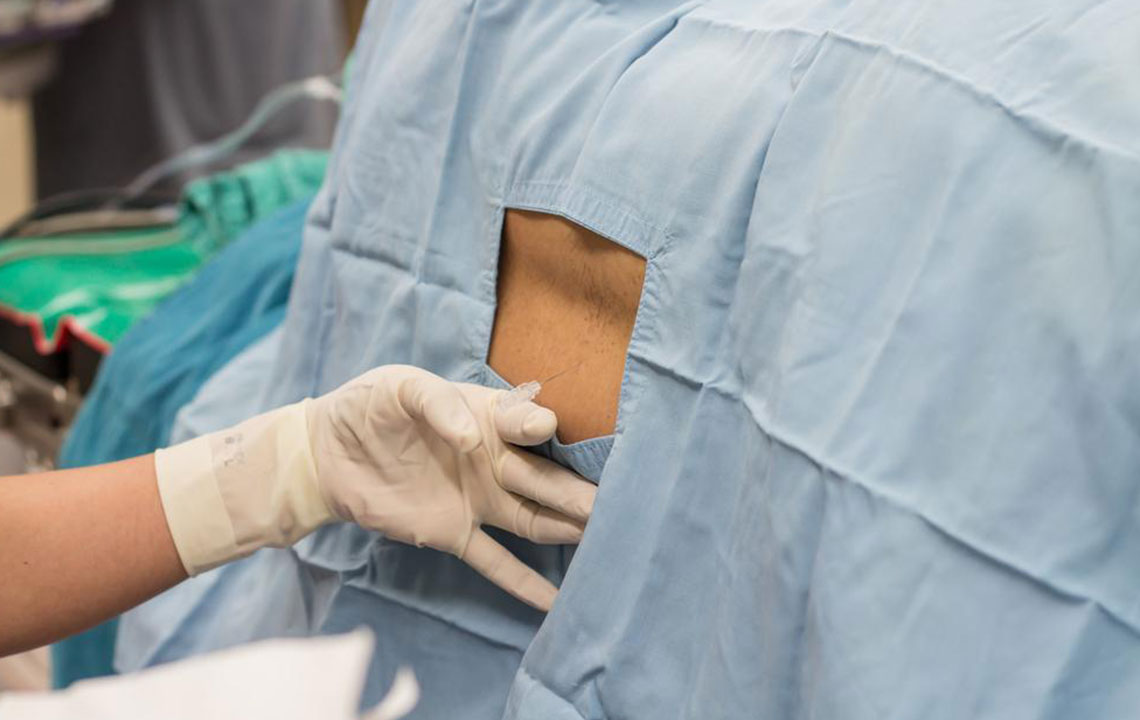
Understanding Ureteroscopy: A Popular Minimally Invasive Treatment
Ureteroscopy is a targeted endoscopic procedure designed to remove or break down kidney stones lodged in the urinary tract. It has gained popularity due to its minimally invasive nature, high success rate, and relatively quick recovery time. The procedure generally lasts about an hour and can be performed under general or spinal anesthesia, ensuring patient comfort throughout.
The surgical process involves several carefully coordinated steps:
This procedure is initiated by inserting a small, flexible endoscope called a cystoscope through the patient's urethra, the natural passage leading to the bladder. Using real-time X-ray imaging for guidance, the surgeon advances the cystoscope into the bladder to visualize and assess the area for any abnormalities.
Once the cystoscope reaches the bladder, a thin guide wire is carefully introduced through the device to serve as a pathway for subsequent instruments. The cystoscope is then gently withdrawn, and a ureteroscope—another specialized thin tube—guides the surgeon directly toward the stone located in the ureter or kidney.
This stepwise approach allows precise targeting of the stone with minimal trauma to surrounding tissues.
Upon reaching the stone, the surgeon has two primary options: either to extract the stone directly using miniature forceps or to fragment it using a laser fiber integrated into the ureteroscope. The laser breaks the stone into tiny pieces, which can then pass naturally through the urinary tract, often reducing the risk of causing further obstruction or discomfort.
After the removal or fragmentation, a temporary ureteral stent or urinary catheter may be placed to facilitate healing and ensure unobstructed urine flow. These are usually removed during follow-up visits after a few days to weeks.
Postoperative Care and Recovery Expectations
Patients are typically observed in the recovery room for at least 24 hours. During this time, medical staff monitor for any immediate complications, such as bleeding or signs of infection, ensuring the patient is stable before discharge.
Resuming daily activities is generally possible within 2 to 3 days, but patients are advised to avoid strenuous exercises and heavy lifting initially. Driving immediately after the procedure is not recommended; arrange for transportation assistance.
Pain management is an important aspect of recovery. Surgeons usually prescribe medications to manage mild to moderate discomfort, especially during urination. Over-the-counter pain relievers are often sufficient.
It’s common for patients to experience slight stinging, burning, or blood in the urine for a few days post-procedure. Drinking plenty of fluids helps flush residual stone fragments and promotes faster recovery.
Follow-up appointments are crucial to assess healing, remove any temporary stents, and evaluate the effectiveness of the treatment. Regular imaging tests may be performed to ensure no new stones are developing.
Potential complications, although rare, include urethral or ureteral injury, infection, or persistent obstruction. Patients should report any severe pain, fever, or difficulty urinating promptly to their healthcare provider.
In conclusion, ureteroscopy represents a significant advancement in the management of kidney stones, combining precision, safety, and efficiency to improve patient outcomes. Its minimally invasive nature means fewer hospital stays, quicker recoveries, and high success rates, making it a preferred choice for both patients and urologists worldwide.