Comprehensive Guide to Early Cancer Detection: Essential Screening Techniques and Innovations
Early cancer detection is vital for successful treatment. This comprehensive guide explores advanced screening methods including imaging techniques like CT, MRI, ultrasound, endoscopy, biopsies, and molecular tests. Learn how these technologies work together to identify cancer at the earliest stages, improving outcomes and survival rates. The article emphasizes the importance of proactive screening and innovations in diagnostic procedures for better patient care.
Comprehensive Guide to Early Cancer Detection: Essential Screening Techniques and Innovations
Early detection of cancer significantly improves treatment outcomes and survival rates. Recognizing the importance of timely diagnosis, healthcare professionals emphasize the use of diverse screening methods that can identify malignant changes before symptoms appear. This proactive approach not only enhances chances for successful intervention but also helps reduce the physical and financial burdens on patients. Various organizations, including the American Cancer Society, have established comprehensive screening guidelines, which serve as a vital resource for both medical practitioners and patients seeking to understand the most effective methods for early cancer detection. Below, we explore key diagnostic procedures and technological advancements that play a crucial role in early cancer diagnosis.
Advanced Imaging Techniques
Imaging modalities remain the cornerstone of early cancer detection, offering non-invasive ways to visualize internal structures, identify abnormal growths, and assess tumor characteristics. These methods provide detailed insights into tumor size, location, and potential spread, allowing clinicians to make informed decisions about further testing or intervention. Various imaging tools are employed depending on the suspected cancer type and location, including ultrasound, X-ray, computed tomography (CT), magnetic resonance imaging (MRI), and nuclear medicine scans. Each technique has unique advantages, and often, a combination of these methods provides the most comprehensive evaluation.
Imaging tests are integral to differentiating benign from malignant tumors, guiding treatment planning, and accurately staging cancer. They also assist in monitoring tumor response post-treatment, ensuring a tailored and effective therapeutic approach. Here’s a detailed look at some prominent imaging techniques:
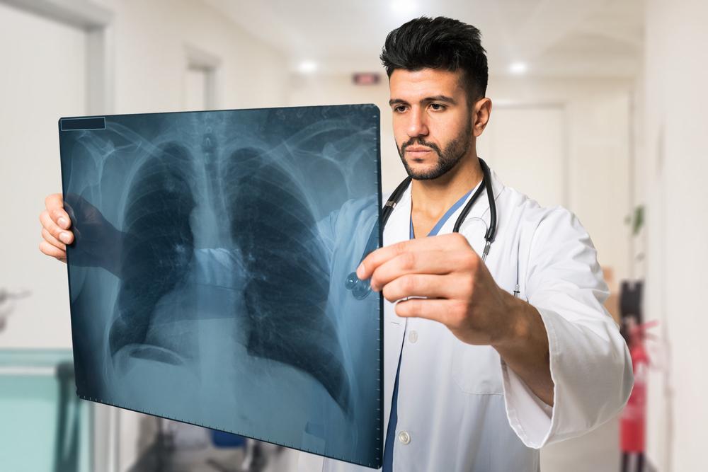
Computed Tomography (CT) Scans
CT scans, also known as CAT scans, are a powerful diagnostic tool that generate cross-sectional images of the body’s bones, organs, and tissues through the use of X-ray technology. These non-invasive and painless procedures provide detailed views, enabling clinicians to locate tumors precisely, measure their size, and evaluate their borders. CT imaging is particularly valuable in diagnosing cancers such as lung, liver, and pancreatic cancers. Additionally, CT-guided procedures, like radiofrequency ablation (RFA), benefit from accurate tumor localization, improving treatment precision.
Magnetic Resonance Imaging (MRI)
MRI employs magnetic fields and radiofrequency waves to produce high-resolution images of soft tissues and internal organs, unlike X-rays, which are better suited for bone imaging. The use of contrast agents like gadolinium enhances soft tissue visualization, facilitating detailed assessment of tumor origin, extent, and invasion into surrounding tissues. MRI is especially valuable in detecting brain, spinal cord, and pelvic cancers, providing critical information for diagnosis and surgical planning. The technique’s non-invasive nature and exceptional tissue contrast make it a preferred choice in many early detection scenarios.
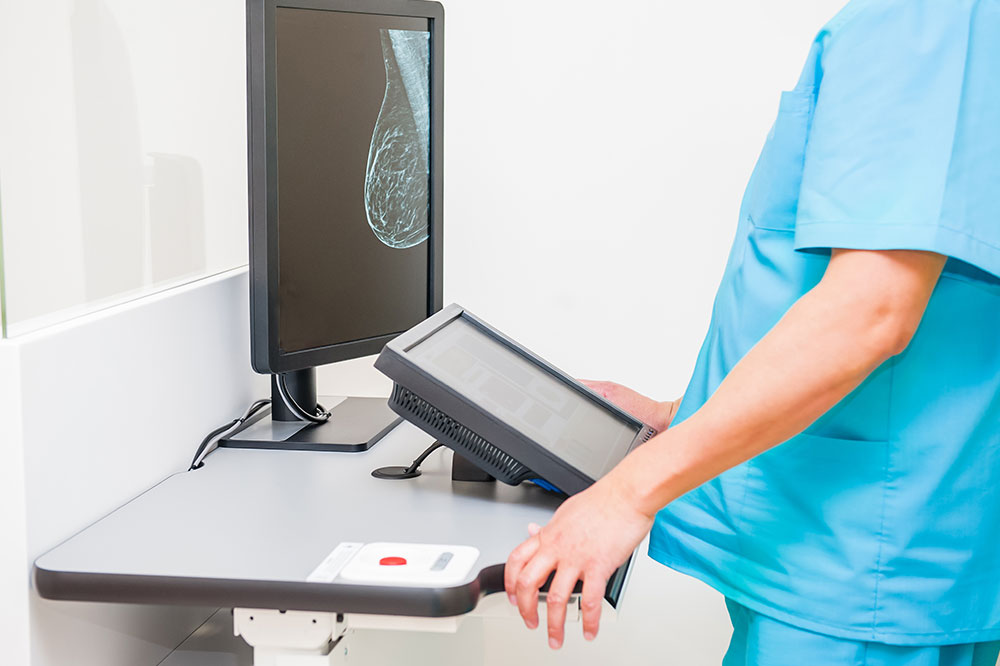
X-ray and Contrast Studies
X-ray imaging remains a fundamental diagnostic method for certain cancers, notably breast cancer, where digital mammography offers highly effective screening and early detection. Contrast-enhanced studies involve the administration of dyes, such as barium or iodine-based agents, to improve visualization of internal structures like the gastrointestinal tract or urinary system. These techniques help delineate tumors, identify blockages, and detect abnormal tissue growths with higher clarity.
Nuclear Medicine Imaging
Nuclear medicine involves the use of radioactive substances, radionuclides, to visualize cellular activity within the body. Techniques such as PET scans (Positron Emission Tomography) detect metabolic changes associated with cancer, highlighting areas of abnormal activity. These scans are particularly useful for staging cancer, assessing treatment response, and detecting metastases. While nuclear scans provide valuable functional information, they are often used in conjunction with other imaging methods for comprehensive diagnosis.
Ultrasound Examination
Ultrasound uses high-frequency sound waves to create real-time images of organs and tissues, making it an invaluable tool for soft tissue evaluation. It’s radiation-free, quick, and effective in distinguishing between cystic (fluid-filled) and solid tumors. Ultrasound is routinely used for assessing thyroid nodules, liver lesions, and ovarian cysts. Its ability to evaluate blood flow with Doppler techniques further enhances its utility in diagnosing vascular abnormalities and tumor vascularity, providing critical insights in early cancer detection.
Endoscopic Procedures
Endoscopy involves inserting a flexible tube with a camera and light into the body to directly visualize internal organs. This minimally invasive technique is essential for detecting cancers within the gastrointestinal tract, such as esophageal, gastric, and colorectal cancers. Procedures like colonoscopy not only allow for direct inspection but also facilitate tissue biopsies, which confirm diagnosis. Endoscopic laser and photodynamic therapies can sometimes be performed during the same procedure to treat superficial tumors, further emphasizing the role of endoscopy in early detection and intervention.
Biopsy and Tissue Sampling
Biopsies are fundamental in diagnosing cancer, involving the removal of tissue samples from suspicious areas for microscopic examination. Techniques vary from simple skin punch biopsies to more complex core needle or surgical biopsies. Fine-needle aspiration biopsy (FNA) is a minimally invasive method used to extract cells from nodules or masses, guiding diagnosis and treatment decisions. Accurate biopsy results are critical for confirming malignancy, determining cancer subtype, and planning appropriate treatment strategies.
Cellular and Fluid Analysis
Analyzing cells and fluids from within the body is a key aspect of early cancer detection. Cytology examines individual cells obtained via scraping, brushing, or fluid collection from sites like urine, respiratory secretions, or cervical smears (Pap tests). These analyses can identify abnormal, precancerous, or malignant cells, providing early warning signs of cancer development. Such tests are often part of routine screenings, aiding in early intervention and improved prognosis.