Comprehensive Overview of the Top 6 Breast Cancer Diagnostic Techniques
This comprehensive article explores the six most effective breast cancer diagnostic methods, including physical exams, mammography, ultrasound, biopsy, MRI, and staging tests. Understanding these techniques is vital for early detection and effective treatment planning, significantly improving patient outcomes. Learn how these tools work together to identify and assess breast cancer at various stages, ensuring timely intervention and better prognosis.

Comprehensive Overview of the Top 6 Breast Cancer Diagnostic Techniques
Breast cancer remains one of the most prevalent and concerning health issues affecting women worldwide, though men are also susceptible. It occurs when abnormal cells in the breast tissue divide uncontrollably, often forming a lump or tumor that can invade surrounding tissues if left undetected. Early detection is crucial for prognosis and treatment efficiency. The development of precise diagnostic methods has significantly improved the ability to identify breast cancer at initial stages, enabling timely intervention and better outcomes. In this detailed overview, we explore the six most effective diagnostic techniques used by healthcare professionals to detect and evaluate breast cancer.
Breast Physical Examination: An essential first step performed by clinicians, this exam involves a thorough inspection and palpation of both breasts and adjacent lymph nodes, particularly those in the underarm (axillary nodes). The physician checks for unusual lumps, thickening, or changes in skin or nipple appearance. Although subjective, this method remains valuable in routine screening and initial assessment, especially in community healthcare settings.
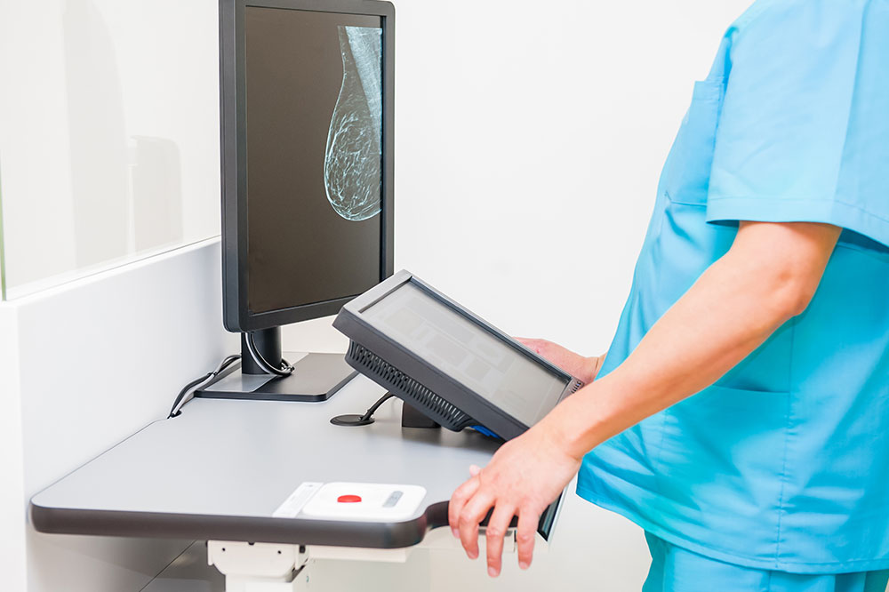
Mammography: Recognized as the gold standard for breast cancer screening, mammography is an X-ray technique that provides detailed images of the breast tissue. It helps detect small tumors or irregularities not palpable during a physical exam. Mammograms can identify microcalcifications—tiny deposits of calcium that may signal early cancer, and are particularly effective in screening women over 40 or at high risk. Conventional 2D mammography is often supplemented with 3D mammography (tomosynthesis) for better visualization.
Ultrasound Imaging: A non-invasive imaging modality that uses high-frequency sound waves to produce real-time images of breast tissue. It is especially useful for distinguishing between solid tumors and benign cysts, guiding biopsy procedures, and evaluating abnormalities detected on mammograms. Ultrasound is safe, free of radiation, and often paired with mammography in diagnostic workups, especially in women with dense breast tissue where mammograms have limitations.
Biopsy: The definitive diagnostic procedure, biopsy involves removing a tissue sample from the suspicious area for microscopic examination. Various techniques exist, including fine-needle aspiration, core needle biopsy, and surgical biopsy. Pathologists analyze the tissue to identify cancer cells, determine tumor type, grade, and hormone receptor status. This information is crucial for planning appropriate treatment strategies and prognosis assessment.
Breast MRI (Magnetic Resonance Imaging): Offering detailed internal images, MRI uses powerful magnets, radio waves, and contrast agents to visualize breast structures. It is especially valuable in high-risk women, dense breast tissue, or for assessing the extent of known tumors. MRI helps detect additional lesions not visible on mammograms or ultrasound and can guide surgical planning. It also plays a role in screening women with genetic predispositions like BRCA mutations.
Staging and Extent Assessment: Once a diagnosis is established, additional imaging studies are conducted to determine how far the cancer has spread. These include blood tests, bone scans, CT scans, PET scans, and MRI. The staging process helps categorize breast cancer from stage 0 (carcinoma in situ) to stage IV (metastatic disease). Accurate staging influences treatment decisions, such as surgery, chemotherapy, radiation, or targeted therapy, and helps predict outcomes.
It's important to note that not every test is necessary for every patient. The choice of diagnostics is tailored based on clinical findings, patient risk factors, and initial imaging results. The comprehensive evaluation aims to detect cancer early, establish the extent of the disease, and facilitate personalized treatment planning. Recognizing the importance of early detection and utilizing the appropriate diagnostic tools can dramatically improve survival rates and quality of life for breast cancer patients.