Ultimate Guide to Melanoma Detection, Diagnosis, and Prevention Strategies
This comprehensive guide covers the importance of early melanoma detection through detailed self-examinations, understanding suspicious signs, and when to seek professional diagnosis. It explains various biopsy techniques used by dermatologists and offers essential prevention tips to reduce skin cancer risks. Early identification and proactive skin health management can significantly enhance treatment success and save lives. Perfect for high-risk individuals or anyone interested in maintaining healthy skin, this article provides everything you need to know about melanoma detection and diagnosis.
Ultimate Guide to Melanoma Detection, Diagnosis, and Prevention Strategies
Recognizing melanoma early is vital for effective treatment and improving survival rates. Melanoma, a type of skin cancer originating in the pigment-producing melanocytes, can develop rapidly if not detected in its initial stages. Therefore, regular skin self-examinations, especially for individuals with high-risk factors, can be life-saving. This comprehensive guide covers how to perform thorough skin checks, identify suspicious lesions, and understand the diagnostic procedures involved in detecting melanoma.
People with fair skin, light hair, a history of frequent sun exposure, or numerous moles and freckles are especially encouraged to monitor their skin closely. Early signs of melanoma often include changes in existing moles or the appearance of new pigmented spots. These changes can manifest as elevation, irregular borders, color variation, flaking, soreness, or bleeding. In this guide, we detail step-by-step instructions for self-examination, when to seek medical advice, and what to expect during professional evaluation.
How to Conduct a Self-Examination
Performing regular skin checks is the first line of defense against melanoma. Use a full-length mirror and a hand-held mirror to examine all areas of your body. Be diligent to assess hidden spots that are often overlooked. Focus on critical areas prone to sun exposure and unique to melanin production:
Back
Shoulders and upper arms
Neck and behind the ears
Chest and abdomen
Scalp (use a mirror or have someone assist)
Face and lips
Both front and back of arms
Groin and pubic area
Legs, including thighs and calves
Feet, including soles and under the toenails
Hands and fingers
During your examination, look for:
Asymmetrical moles or spots
Irregular borders that are ragged or blurred
Multiple colors within a single lesion, such as brown, black, red, white, or blue
Evolution or noticeable changes in size, shape, or color of a mole over time
New growths or spots that look different from your other moles
When to See a Dermatologist
If you notice any of the signs mentioned above, or if a mole looks unusual or changing, schedule an appointment with a dermatologist promptly. Early diagnosis drastically increases the chances of successful treatment. Additionally, individuals with risk factors such as fair skin, a family history of melanoma, a high number of moles, or previous sunburns should consider regular professional skin evaluations, typically once a year.
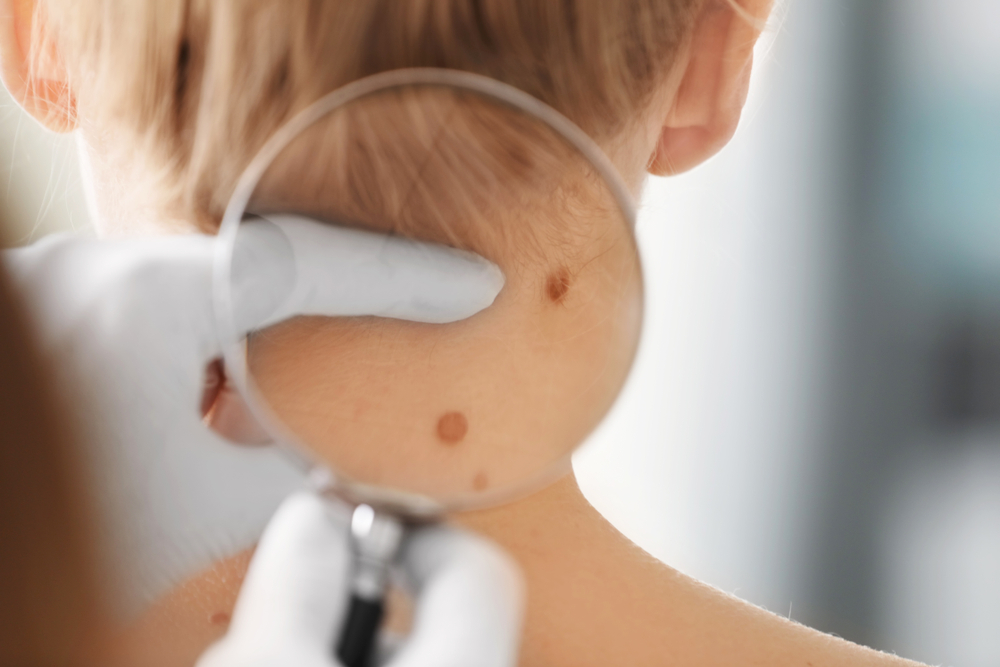
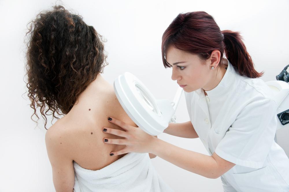
Dermatologists are trained to perform detailed skin assessments and can identify subtle changes unnoticed by the untrained eye. They may utilize advanced tools such as dermoscopy or digital mole monitoring systems to detect early melanomas. Regular checkups are especially recommended for high-risk populations.
Diagnostic Procedures for Melanoma
When a suspicious lesion is identified, a dermatologist will recommend diagnostic procedures to confirm whether it is melanoma. The most common method is a skin biopsy, which involves removing a sample of tissue for laboratory analysis. Various biopsy techniques are employed based on the lesion's location, size, and appearance.
Types of Biopsies
Punch Biopsy: Utilizes a circular blade to extract a small, round tissue sample, capturing all skin layers. This method is ideal for examining thicker or irregular pigmented lesions.
Optical Biopsy: A non-invasive imaging technology known as reflectance confocal microscopy (RCM) allows for real-time visualization of skin layers without cutting. While not a replacement for histopathology, it aids in evaluating ambiguous lesions.
Shave (Tangential) Biopsy: Involves shaving off the top layers of skin using a surgical blade. Suitable for flat or raised lesions, though it may require bleeding control measures.
Fine Needle Aspiration (FNA): Uses a thin needle guided by ultrasound or other imaging techniques to extract cells from deeper lesions or lymph nodes suspected of harboring metastasis. This procedure assists in staging cancer spread.
Incisional Biopsy: Removes a portion of a large lesion, especially when the lesion cannot be entirely excised in one procedure. The sample undergoes histological examination for definitive diagnosis.
Sentinel Lymph Node Biopsy: Particularly in invasive melanoma, this involves injecting a dye or radioactive tracer to identify the first lymph node(s) draining from the tumor. Removal and analysis of these nodes help determine metastasis and staging.
Following biopsy confirmation of melanoma, treatment options may include surgical excision, immunotherapy, targeted therapy, chemotherapy, or radiation, depending on the stage of the disease. Early diagnosis is crucial for a positive prognosis.
Prevention and Risk Reduction
In addition to regular self-examinations and professional screenings, adopting sun safety habits reduces melanoma risk. This includes:
Applying broad-spectrum sunscreen with SPF 30 or higher daily, even on cloudy days
Wearing protective clothing, hats, and UV-blocking sunglasses
Seeking shade during peak sunlight hours (10 a.m. to 4 p.m.)
Avoiding tanning beds and artificial UV exposure
Performing regular skin checks and staying vigilant for any changes
By incorporating these precautions, individuals can significantly decrease their risk of developing melanoma and other skin cancers.
Conclusion
Early detection of melanoma through self-examination and prompt medical evaluation can save lives. Understanding how to identify suspicious signs and knowing when to seek professional help are essential steps in skin health. Remember, if you notice any unusual changes in your skin, consult a dermatologist without delay. Regular skin assessments are a proactive way to catch melanoma early and improve treatment outcomes.