Effective Approaches for Managing and Treating Fuchs Endothelial Corneal Dystrophy
This comprehensive guide explores effective management strategies for Fuchs Endothelial Corneal Dystrophy, covering early symptoms, non-invasive treatments, surgical options, and future research directions. It emphasizes early detection and personalized care to preserve vision and improve patient outcomes through the latest advancements in eye treatment technology.

Effective Approaches for Managing and Treating Fuchs Endothelial Corneal Dystrophy
Fuchs Endothelial Corneal Dystrophy (FECD) is a progressive degenerative eye condition primarily affecting the corneal endothelium—the innermost layer of the cornea responsible for maintaining corneal clarity by regulating fluid balance. This disorder gradually impairs the function of these vital endothelial cells, leading to fluid accumulation, corneal swelling, cloudiness, and ultimately, a decline in visual acuity. Understanding the disease mechanism, early symptoms, and the spectrum of available management techniques is crucial for patients and healthcare providers striving to preserve vision and improve quality of life.
Understanding Fuchs Endothelial Corneal Dystrophy
FECD typically manifests in individuals over the age of 50, although early signs can sometimes be detected in younger adults. The condition begins subtly, with the endothelial cells gradually deteriorating, which diminishes their ability to maintain the cornea's dehydration status. As endothelial cell health declines, patients experience corneal edema—swelling caused by fluid retention—leading to visual disturbances and discomfort.
Early diagnosis is vital to slow the disease's progression and prevent severe visual impairment. The primary symptoms of FECD include blurred vision, especially in the morning when corneal swelling is at its peak, halos and glare around lights, ocular soreness or irritation, and increased sensitivity to light (photophobia). Recognizing these symptoms early enables patients to seek timely medical advice, potentially avoiding more invasive treatments later.
Understanding the pathophysiology of FECD helps inform treatment options that range from conservative management to surgical intervention. Each approach aims to alleviate symptoms, reduce disease progression, and restore or preserve visual function.
Non-Invasive Management Strategies
Despite the absence of a definitive cure for FECD, several non-invasive or conservative treatments can significantly improve patient comfort and visual clarity. These strategies are especially crucial during early or mild stages of the disease and often serve as adjuncts or palliative measures alongside surgical options.
1. Artificial Tears and Lubricants
One of the simplest and most accessible treatments involves the use of over-the-counter artificial tears. These lubricating eye drops help maintain moisture on the corneal surface, alleviating dryness and soreness that often accompany FECD. While they do not directly address the underlying endothelial cell degeneration, they provide vital symptomatic relief, especially during the day or in environments with low humidity. Regular use can help sustain comfort and prevent secondary irritation caused by dry eyes.
2. Hypertonic Saline Solutions
For managing corneal edema, hypertonic saline solutions—typically 5% saline drops or ointments—are highly effective. These solutions work by drawing excess fluid out of the cornea through osmotic principles, reducing swelling and restoring better visual acuity. Patients are generally advised to use these treatments in the morning when swelling tends to be most prominent. Consistent use can temporarily improve vision and alleviate discomfort caused by fluid buildup.
3. Home-Based Dehydration Techniques
Innovative homemade interventions can aid in corneal dehydration, especially for symptomatic relief. One such technique involves using a warm or cool setting on a hairdryer held at arm's length directed toward the eyes for a few minutes daily. This method aims to promote evaporation of excess surface fluid on the cornea, thereby reducing swelling and improving clarity. Patients should exercise caution to avoid direct contact with high heat levels that could harm the eyes.
In addition to these traditional strategies, lifestyle modifications such as wearing UV-protective sunglasses, managing environmental factors like humidity, and avoiding eye irritants can contribute to overall eye health and comfort.
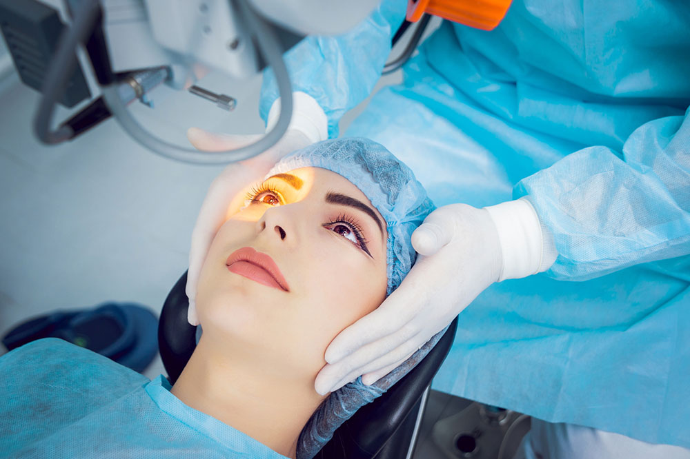
Surgical Treatment Options
As FECD advances, conservative treatments may become insufficient to control symptoms or halt disease progression. In such cases, surgical intervention becomes necessary to restore corneal clarity and function. Several surgical techniques are available, each tailored to the severity of the condition and the patient's specific needs.
1. Endothelial Keratoplasty (EK)
This minimally invasive procedure involves replacing the diseased endothelial layer with healthy donor tissue. It has become the preferred surgical method due to its faster recovery times and better visual outcomes compared to traditional full-thickness transplants. Types include:
— DSEK (Descemet Membrane Endothelial Keratoplasty): This procedure transplants both the Descemet membrane and the endothelial cell layer. It is widely used and offers good outcomes with relatively quick recovery.
— DMEK (Descemet Membrane Endothelial Keratoplasty): This more advanced form transplants only the Descemet membrane and endothelium, resulting in a thinner graft and often sharper vision. DMEK is gaining popularity for its rapid visual recovery and reduced rejection rates.
2. Full-thickness Corneal Transplantation
Penetrating Keratoplasty (PK) involves replacing the entire cornea with a donor graft. Although it is less commonly performed now due to the success of EK procedures, it remains an option in cases of extensive scarring or other corneal pathologies. PK requires a longer recovery period and carries higher risks of complications such as rejection and graft failure.
3. Managing Graft Rejection and Complications
Postoperative care is critical for graft success. Early detection of signs such as redness, pain, photophobia, or declining vision allows for prompt treatment to prevent graft rejection. This often involves the use of anti-rejection medications and regular follow-up appointments with ophthalmologists to monitor graft health.
Future Directions and Innovative Treatments
The field of FECD management is dynamic, with ongoing research focusing on novel methods to delay or reverse disease progression. Recent advancements include:
— The use of femtosecond laser technology enhances surgical precision during endothelial transplants, leading to better outcomes.
— Pharmacological research aims to develop drugs that support endothelial cell health and function, potentially reducing the need for immediate surgery.
— Cutting-edge approaches like gene therapy and regenerative techniques hold promise for repairing or replacing defective endothelial cells at the genetic level, paving the way for future cures.
While challenges remain, continual scientific progress offers hope that FECD can be managed more effectively in the future. Early diagnosis, personalized treatment plans, and ongoing advancements in eye surgery and regenerative medicine are central to improving patient outcomes.
Patients are encouraged to consult specially trained ophthalmologists promptly and stay informed about emerging treatment options. Active participation in disease management can significantly influence the preservation of vision and overall quality of life.