Comprehensive Overview of Pulmonary Embolism: Causes, Symptoms, Diagnosis, and Effective Treatments
This comprehensive article explores pulmonary embolism, detailing its causes, symptoms, diagnostic procedures, and treatment options. It emphasizes the importance of early detection, understanding risk factors, and preventive measures. Readers will gain insights into how blood clots originate from deep veins, the signs indicating PE, and the latest medical approaches to manage this life-threatening condition effectively. Enhanced awareness can lead to timely intervention, potentially saving lives and reducing complications associated with pulmonary embolism.

Deep Dive into Pulmonary Embolism: Causes, Signs, Diagnostic Methods, and Treatment Options
Pulmonary embolism (PE) represents a serious medical emergency that arises when a blood clot blocks an artery in the lungs. This condition often originates from deep vein thrombosis (DVT) in the legs, where blood clots form and then dislodge, traveling through the bloodstream to lodge in the pulmonary arteries. Although less common, PE can also occur due to other embolic materials such as fat, air bubbles, or tumor fragments blocking lung arteries. Timely diagnosis and intervention are critically important because PE can quickly become life-threatening by impairing oxygen exchange and causing cardiovascular instability.
Understanding the risk factors, recognizing symptoms early, and knowing the diagnostic processes can significantly improve outcomes. Prevention strategies mainly involve reducing factors that contribute to abnormal blood clot formation, particularly in the lower extremities, thereby reducing the incidence of pulmonary embolism and its severe complications.
Recognizing the Symptoms of Pulmonary Embolism
The clinical presentation of PE varies depending on the size of the clot, the extent of lung area affected, and the individual's overall health. Common manifestations include abrupt onset of shortness of breath that worsens with exertion, which is often the most prominent symptom. Chest pain associated with PE can mimic that of a heart attack, described as sharp, stabbing, or pleuritic pain that worsens with deep breaths or coughing. Coughing may produce hemoptysis, or blood-streaked sputum, especially in more severe cases. Patients often report dizziness or fainting episodes, rapid heartbeat (tachycardia), profuse sweating, and a feeling of anxiety or dread.
Additionally, skin may appear blue or discolored (cyanosis) if oxygen levels plummet. Swelling, warmth, tenderness, or pain in the legs—especially in the calves—may indicate underlying DVT, the source of many emboli. Patients with pre-existing heart or lung conditions may experience more pronounced symptoms or atypical presentations, making diagnosis more challenging.
Major Causes of Pulmonary Embolism
Pulmonary embolism predominantly results from emboli originating in the deep veins of the legs or pelvis, known as deep vein thrombosis (DVT). When clots form in these veins, they may detach and travel through the venous system towards the lungs, where they lodge in pulmonary arteries. Other less common sources include emboli composed of fat from broken bones, air bubbles introduced during medical procedures, or tumor fragments from cancers like lung, ovarian, or pancreatic cancers. These events impair blood flow in the lungs, which can lead to reduced oxygen delivery and, if untreated, result in pulmonary infarction—a condition where lung tissue dies due to oxygen deprivation. In severe cases, this can cause sudden respiratory failure and death.
Factors That Increase the Risk of Developing Pulmonary Embolism
While anyone can develop blood clots under certain circumstances, specific risk factors significantly elevate the likelihood. A personal or family history of clotting disorders predisposes individuals to thrombosis. Cardiovascular diseases, such as atrial fibrillation or coronary artery disease, heighten the risk by affecting blood flow and clotting mechanisms. Certain cancers—particularly lung, ovarian, pancreatic, and gastrointestinal—are associated with hypercoagulability, increasing clot formation. Additionally, treatments like chemotherapy can damage blood vessels or alter clotting factors, further raising risk.
Other important risk factors include recent major surgeries, especially orthopedic procedures involving hips or knees, prolonged immobility such as extended bed rest or long-haul flights, smoking, obesity, pregnancy, and hormone therapy with estrogen or oral contraceptives. Recognizing these risk factors enables healthcare providers to implement preventive strategies, including prophylactic blood thinners and lifestyle modifications, to reduce PE incidence.
Diagnostic Approaches for Pulmonary Embolism
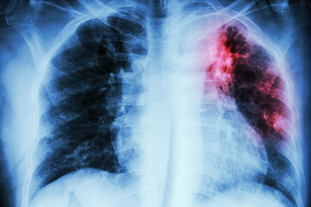
Detecting PE requires a combination of clinical assessment and sophisticated imaging techniques. Physicians begin by evaluating symptoms, medical history, and physical examination findings to estimate pre-test probability. Diagnostic tests include chest radiography (X-ray) to rule out other causes of symptoms and assess lung status. Electrocardiograms (ECGs) help exclude heart-related issues. Advanced imaging modalities such as computed tomography pulmonary angiography (CTPA) are gold standards for visualizing clots within pulmonary arteries. Magnetic resonance imaging (MRI) may be used in certain cases. Pulmonary angiography, an invasive but definitive test, involves injecting contrast dye into lung vessels to identify blockages.
Blood tests like D-dimer are useful screening tools since elevated levels suggest active clot formation, but they are not definitive alone. Duplex ultrasound of the legs is performed to detect DVT, which often propagates to cause PE. Combining clinical scoring systems, such as the Wells score, with these diagnostic tools helps determine the need for further testing and ensures prompt diagnosis.
Effective Treatment Strategies for Pulmonary Embolism
Management of PE aims to restore normal blood flow, prevent clot propagation, and reduce the risk of recurrence. Early treatment typically involves anticoagulant medications, such as heparin, warfarin, or direct oral anticoagulants (DOACs), to thin the blood and prevent new clots from forming. In cases where the clot is large or causing hemodynamic instability, thrombolytic therapy—using drugs like alteplase—may be administered to dissolve the clot rapidly.
Severe or life-threatening PE might necessitate surgical intervention, such as embolectomy, to physically remove the clot. In some cases, inserting an inferior vena cava (IVC) filter—a device placed in the large vein that returns blood to the heart—can trap emboli before reaching the lungs, particularly in patients unable to tolerate anticoagulation.
Long-term management focuses on preventing recurrence through continued anticoagulation therapy, use of compression stockings to promote blood flow, and leg exercises to reduce stasis. Identifying and managing underlying risk factors are critical components of comprehensive care. Patient education about recognizing symptoms of recurrent PE and adhering to treatment plans enhances prognosis and quality of life.


