Comprehensive Guide to PET Imaging in Lung Cancer Detection and Progression Evaluation
PET imaging plays a vital role in detecting lung cancer and assessing its progression. By combining metabolic and anatomical data, PET scans enable precise diagnosis, staging, and monitoring of treatment response. This advanced technology aids clinicians in differentiating malignant tissues from benign, planning effective treatment strategies, and improving patient outcomes. Its minimally invasive approach and detailed results make PET an indispensable tool in lung cancer management, ensuring timely and accurate intervention for better prognosis.
Comprehensive Role of PET Imaging in Detecting and Monitoring Lung Cancer Progression
Positron emission tomography (PET) has revolutionized medical imaging, offering advanced insights into organ function at the cellular level. Unlike traditional imaging systems that focus primarily on anatomy, PET scans utilize radioactive tracers to visualize metabolic processes within tissues. This capability makes PET particularly valuable in the diagnosis, staging, and management of various cancers, especially lung cancer. When combined with computed tomography (CT), PET provides a hybrid imaging modality that delivers highly detailed, three-dimensional images, enabling clinicians to distinguish between healthy and abnormal tissue with remarkable precision.
In the context of lung health, PET imaging is often paired with CT scans to improve diagnostic accuracy. Patients presenting symptoms such as persistent cough, unexplained weight loss, chest pain, or coughing up blood are typically recommended for both imaging tests. This combined approach enhances the ability of physicians to obtain comprehensive views of the lungs, making it easier to identify malignant tumors, differentiate them from inflammatory processes, and assess their extent.
How PET Imaging is Used During Lung Cancer Diagnosis
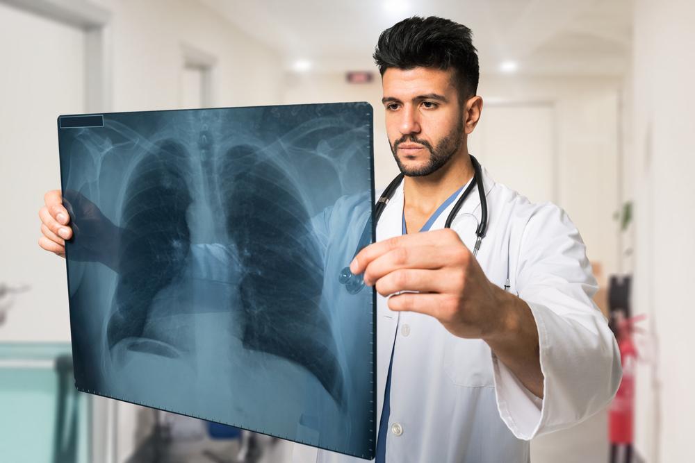
The diagnostic process begins with the patient receiving an injection of a radiotracer—most commonly glucose tagged with a radioactive isotope—about an hour prior to the scan. Since cancer cells tend to consume glucose at a higher rate than normal cells, they absorb more of the radioactive tracer. This increased uptake signifies areas of heightened metabolic activity, often corresponding to tumor sites.
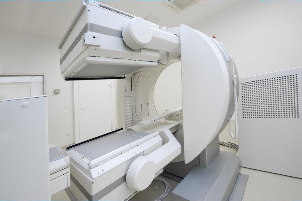
Once injected, the patient lies comfortably within the PET scanner, a device that detects gamma rays emitted when the radioactive tracer decays. These emissions are recorded and processed by sophisticated computer algorithms to generate detailed images, highlighting regions of abnormal cellular activity. The entire procedure is non-invasive and minimally discomforting, primarily involving a brief injection that may cause a slight pinch or sting. The length of the scan varies but generally lasts between 30 to 60 minutes, depending on the targeted area.
Using PET Scans to Determine Lung Cancer Stage
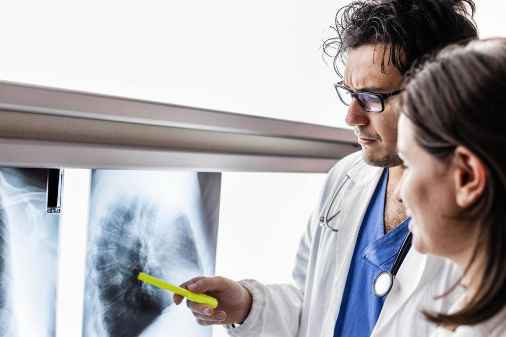
PET imaging is instrumental in accurately staging lung cancer, which is crucial for determining the appropriate treatment approach. The staging process involves assessing how far the cancer has spread within the lung and to other parts of the body. Tissue regions that absorb more radiotracer typically indicate more aggressive or rapidly dividing cancer cells. Thus, PET scans can detect primary tumors and distant metastases that might not be visible on conventional imaging modalities.For example, a stage I tumor indicates localized disease confined to the lung, while stage IV reveals distant metastasis, such as to the brain or bones. Rapidly growing tumors often show higher radiotracer uptake, and this metabolic information helps clinicians assess the aggressiveness of the disease. Importantly, PET scans can also identify small metastatic deposits that are crucial for accurate staging but may be missed by CT or MRI alone.
This comprehensive staging aids in prognosis and guides therapy choices, including surgery, chemotherapy, targeted therapy, or immunotherapy. Moreover, PET scans are also used during treatment to evaluate response, ensuring that therapy is effectively reducing tumor activity or identifying early signs of recurrence.
The entire PET imaging process is a highly valuable diagnostic tool because it combines functional and structural data, providing a holistic overview of lung cancer progression. Its minimally invasive nature, coupled with its high diagnostic accuracy, makes it a cornerstone in modern oncological imaging.