Comprehensive Guide to Bone Mineral Density Testing: Techniques, Importance, and What You Need to Know
This comprehensive guide explores bone mineral density (BMD) testing methods like DXA and QCT, emphasizing their importance in diagnosing osteoporosis early. It explains result interpretation with T-scores and Z-scores, highlighting preventive care strategies. Early detection through BMD testing helps reduce fracture risk and improve quality of life, making it essential for at-risk individuals. The article provides detailed insights into procedures, preparation, and clinical applications, offering valuable knowledge for patients and healthcare providers seeking effective bone health assessment.

Comprehensive Guide to Bone Mineral Density Testing: Techniques, Importance, and What You Need to Know
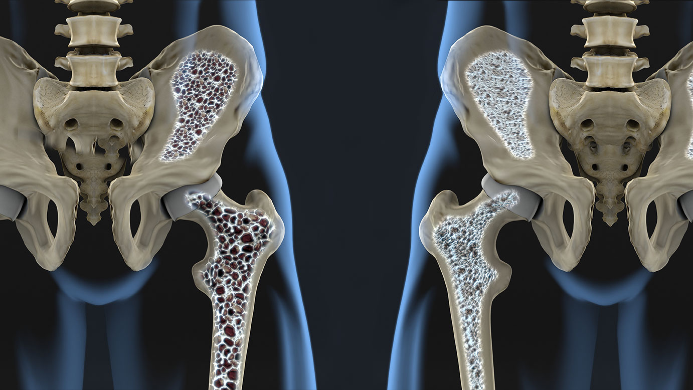
Monitoring bone health is a critical component in diagnosing and managing osteoporosis, a condition characterized by weakened bones and an increased risk of fractures. Bone mineral density (BMD) testing stands out as a vital diagnostic tool that provides precise measurements of bone strength and quality. Traditionally, osteoporosis was only diagnosed after a fracture occurred, which often meant irreversible damage and limited treatment options. However, advances in medical imaging have made it possible to detect bone loss early, allowing for timely intervention and better disease management.
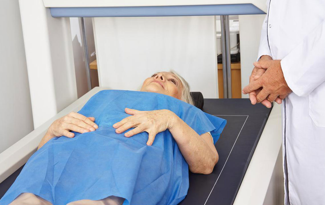
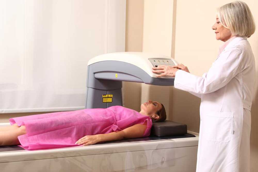
Bone mineral density testing is akin to an advanced form of X-ray imaging. During the procedure, the patient lies on a specialized platform while a small mechanical arm moves over specific bones, primarily focusing on critical areas such as the spine and hips. This process is entirely safe, uses very low doses of radiation, and typically lasts between 10 and 30 minutes, making it a convenient option for routine screening, especially for those at higher risk of osteoporosis.
Methods of Bone Mineral Density Testing
The two most common methods employed for BMD assessment are Dual-energy X-ray Absorptiometry (DXA or DEXA) and Quantitative Computed Tomography (QCT). Each has its specific applications, advantages, and limitations, allowing healthcare providers to choose the most suitable option based on individual patient needs.
Dual-energy X-ray Absorptiometry (DXA)
The DXA scan is the most widely used and recognized method for measuring bone mineral density. It involves minimal radiation exposure, making it a safe and efficient procedure. During a DXA scan, the machine uses two X-ray beams at different energy levels to scan the targeted bones. The differential absorption of X-rays helps calculate the bone mineral content and density. This method provides highly accurate results that are essential for diagnosing osteoporosis and assessing fracture risk.
Preparing for a DXA scan is straightforward—patients should avoid wearing jewelry, belts, or metal objects that could interfere with the scan’s accuracy. Moreover, it’s recommended to refrain from calcium and vitamin D supplements for at least 24 hours prior to the test to avoid skewed results. The procedure itself is painless and quick, offering results that guide treatment decisions effectively.
Quantitative Computed Tomography (QCT)
QCT utilizes a standard CT scanner to analyze bone density, especially useful in cases where spinal deformities like scoliosis or surgical hardware may interfere with traditional DXA scans. Compared to DXA, QCT provides three-dimensional imaging, allowing for a detailed assessment of bones and surrounding soft tissues. It’s particularly beneficial in measuring trabecular (spongy) bone, which tends to deteriorate earlier in osteoporosis.
QCT results are quantitative and can be more sensitive in certain patient populations. It offers a faster turnaround for results and can produce volumetric density measurements, providing a comprehensive picture of bone health. However, due to higher radiation exposure compared to DXA, QCT is typically used selectively for specific clinical situations rather than routine screening.
Understanding BMD Test Results
Interpreting the results of BMD tests involves two key scores: T-score and Z-score. These scores help healthcare providers determine the extent of bone loss and identify the presence of osteoporosis or osteopenia.
What is a T-score?
The T-score compares an individual's bone mineral density to the average peak bone density of a healthy young adult of the same sex. It essentially indicates how much your bones have weakened or remained strong relative to youthful, healthy bones. A T-score of -1. or higher suggests normal bone density. Scores between -1 and -2.5 indicate low bone mass, or osteopenia. A T-score of -2.5 or lower confirms the diagnosis of osteoporosis, reflecting significant bone loss that warrants treatment and lifestyle modifications.
What is a Z-score?
The Z-score compares your bone mineral density to those of your peers of the same age, sex, and ethnicity. A Z-score below -2. suggests that factors other than aging—such as medication effects, nutritional deficiencies, or underlying diseases—may be contributing to abnormal bone loss. This score helps clinicians investigate potential secondary causes of osteoporosis in younger individuals or those without typical risk factors.
Regular BMD testing and careful interpretation of the results are essential for early diagnosis and management of osteoporosis. With early detection, patients can adopt lifestyle changes, dietary adjustments, or medications to prevent fractures and maintain quality of life.
In summary, bone mineral density testing is a vital tool in the proactive management of bone health. Whether via DXA or QCT, these tests provide crucial insights into bone strength, enabling physicians to intervene before fractures occur, thus improving long-term health outcomes for at-risk populations.